Feet

Pressure ulcer categories

The following text is a direct transcript from “Prevention and Treatment of Pressure Ulcers: Short Version of Guidelines 2019”.

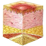
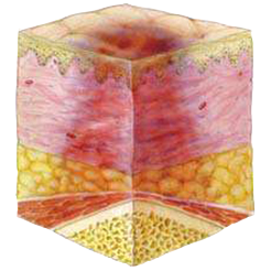
Category I – Redness that does not reduce on pressure

Intact skin with redness in a clearly defined area, usually above a bone protuberance, that does not reduce on pressure. Dark, pigmented skin might not show this sign, though the colour is different from the surrounding areas of skin. The area may be painful, solid or soft, and warmer or colder than other areas of skin. Category I pressure ulcers may be difficult to detect in people with dark skin tones. Category I pressure ulcers may be a sign that the patient is in the risk zone for developing deeper pressure ulcers (1).


Category II – Partial skin damage

Partial skin damage that appears as a superficial open ulcer with a pinkish-red wound bed without fibrin slough. It may also be an intact or open/ruptured serum-filled or blood-filled blister. Appears as a shiny or dry superficial ulcer without fibrin slough or superficial haematoma. This category should not be used to describe skin tears, tape burns, incontinence-associated dermatitis or maceration.(1)

Category III – Full skin damage

Full skin damage. Subcutaneous fat is visible, though not bone, tendon or muscle. Fibrin slough may be visible, though without obscuring the depth of the injury. May include undermining and tunnelling. The depth of a Category III ulcer may vary depending on its anatomical location. The bridge of the nose, ears, back of the head and ankles do not have any subcutaneous fatty tissue and Category III pressure ulcers may be superficial at these locations. In contrast, areas of significant subcutaneous fatty tissue can develop extremely deep Category III pressure ulcers. Bone/tendon is not visible or palpable.(1)

Category IV – Deep full-tissue damage

Deep full-tissue damage involving bone, tendon or muscle. There may be visible fibrin or necrosis. There is often under-mining and tunnelling. The depth of Category IV pressure ulcers varies depending on anatomical location. For example, there is no subcutaneous fatty tissue at the bridge of the nose, ears, back of the head or ankles, and ulcers at these locations may be superficial.

Category/grade IV pressure ulcers may involve muscles and supporting structures (e.g. fascia, tendons or joint capsules), which means that osteomyelitis and osteitis may occur. Exposed bone and muscle is visible or directly palpable. Black necrosis is assessed as category/grade 4 even if the skin is intact, as may be the case with the heels, for example.(1)

Unclassifiable Pressure Ulcer: Depth Unknown

Full-thickness skin damage where the base of the wound is obscured by a fibrin coating (yellow, yellow-brown, gray, green, or brown) and/or necrosis (yellow-brown, brown, or black). Until a sufficient amount of fibrin and/or necrosis is removed, the true depth, i.e., Category/Stage, cannot be assessed. Stable necrosis (dry, firm, intact without redness or fluctuation) on heels serves as the “body’s natural (biological) ‘bandage’” and should not be removed (1).

Suspected Deep Tissue Injury: Depth of the wound unknown

Purple or maroon discoloration of intact skin and/or a blood-filled blister caused by damage to underlying tissue due to pressure and/or shear. The area may be preceded by the skin being painful, firm, spongy, warmer, or cooler compared to the surrounding tissue.

Deep tissue injury can be challenging to detect in individuals with dark skin tones. The progression may involve the formation of a thin blister over a dark wound bed. The wound may continue to progress and become covered by a thin layer of necrosis. Despite optimal treatment, the progression can rapidly involve additional tissue layers.(1)

How can we prevent pressure ulcers? Read more

References
(1) European Pressure Ulcer Advisory Panel, National Pressure Injury Advisory Panel and Pan Pacific Pressure Injury Alliance. Prevention and Treatment of Pressure Ulcers/Injuries: Clinical Practice Guideline. The international Guideline. Emily Haesler (Ed.). EPUAP/NPIAP/PPPIA: 2019